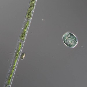
I took some students out to our local lake, Lake Artemesia, and did a couple of plankton tows and a “weed squeeze”. While looking at the samples I took this micrograph and then realized after the fact that it encapsulates much of plastid evolution in one photo.
The filament on the left is Mougeotia, a member of the Zygnematales (the conjugating green algae) and a close relative of land plants. It was used in the 1970s as a model system for classic studies of phytochrome responses. If you shine bright light on the cell the chloroplast will rotate edge-on, presumably to reduce photodamage to the plastid. If you narrow that beam of light down so that it only illuminates one portion of the plastid, that part of the plastid will rotate, but not the portions that remained in dim light. And it is possible to measure the action spectrum of that reaction; it matches the action spectrum for phytochrome, a key plant light sensor. Phytochrome is thought to be fairly widespread among the algae, but now that it is understood that the zygnematophytes are among the closest living relatives of plants, it might be a good time to resume their use as model systems.
The cryptomonad is too small to see really clearly in the micrograph, but it is an example of a lineage that has secondary plastids derived from red algae. In cryptomonads the secondary nature of the plastid is quite obvious (once you understand what is going on) because there is a second, highly reduced, eukaryotic nucleus that is in a separate membrane-bound compartment along with the plastid, and contains genes that can be shown to be of red algal origin. There was a really nice article by Curtis et al. (2012;doi:10.1038/nature11681) that explored the genome of the cryptomonad Guillardia and compared it to Chlorarachnion, a second organism with secondary plastids, although in that case from a green alga.
To the right is Glaucocystis, probably Glaucocystis nostochinearum, although I would be hard pressed to tell you what other species of Glaucocystis there might be. What is interesting about Glaucocystis is that it has plastids that look very much like free-living cyanobacteria. In fact, they even have bacteria-like peptidoglycan cell walls, although these are quite thin when compared to typical cyanobacterial cell walls. These structures seemed so much like cyanobacteria that they were even given their own genus name, but they also seemed very much like organelles, and were never found living outside of Glaucocystis. Again, with the clarity of hindsight we can say it all makes sense — plastids are endosymbiotic cyanobacteria, so the plastids of Glaucocystis — like all other plastids — are both cyanobacteria and organelles. These plastids do have the highly reduced genomes characteristic of other plastids, and glaucocystophytes are probably monophyletic with green and red algae.

